Infrared Microspectroscopy
Performance of the IR microscope is greatly enhanced with infrared synchrotron radiation, which has 100-1000 times higher brightness than a conventional thermal source, makes possible to investigate the chemical variations across the small size samples and also small heterogeneous regions of large samples with high spatial resolution. A small area of 30 micron x 30 micron of the sample can be analyzed with the FT-IR spectrometer and the IR microscope.

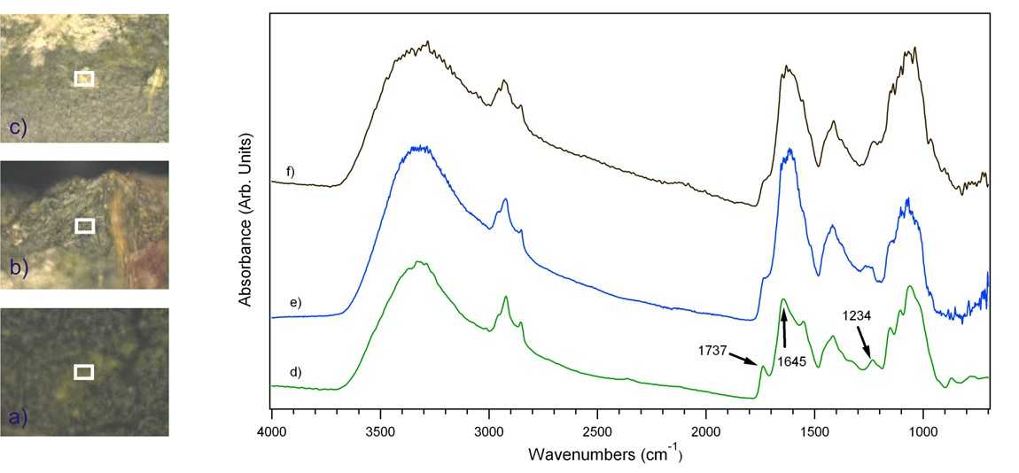
The host-pathogen interaction of bean rust Uromyces appendiculatus with bean leaves has been characterized with the IR microspectroscopy. The optical image of a healthy bean leaf (a), less infected (b), and highly infected (c) parts of an infected bean leaf are shown in the left. The transmission mode with 40x30 μm2 aperture size (the rectangle box in the optical images) was employed for the collection of IR spectra of these samples. IR spectra acquired from healthy, less and highly infected samples are plotted in the right figure, respectively. Evident changes are elucidated in the features of proteins (~1650 cm-1) and carbohydrates (~1100 cm-1). A well resolved protein Amide I band at 1645 cm-1 becomes very broad peak in the IR spectra of infected leaf. This change may arise as a result of differences in the protein structure of healthy and infected bean leaves. Data measured at the CAMD IR Microspectroscopy Beamline.
Supported beamline
- Infrared Microspectroscopy beamline
Click for Beamline Information
Research/Technical Contact: Orhan Kizilkaya